Particle image velocimetry (PIV)

We have validated a novel procedure of using particle image velocimetry (PIV) as a method to determine performance differences between various endovascular catheters (i.e. aspiration catheter systems).
- PIV can help quantify and statistically distinguish differences in applied stress and strain fields between the various catheter types
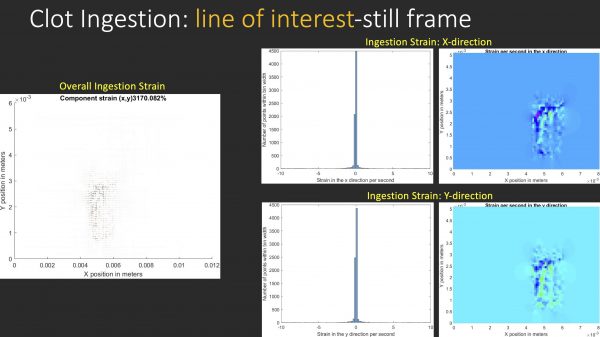
- Flow or clot ingestion can be captured with high-speed cameras utilizing laser fluorescence of the PIV particles within the blood analog and synthetic clots, along with shadowgraph images
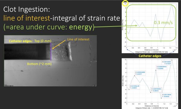
- Resulting data can be used to create velocity vector position, strain rate, and stress graphs of clot ingestion by both catheter types
- Multiplying strain rate by the compression and shear modulus of the clots provides overall stress values of clot ingestion.
- The area under a strain rate curve provides the energy efficiency of the catheter system
- Stress is proportional to overall aspiration force at the tip (Force = stress/catheter tip area)
Clot diameters were optimized, based on catheter tip diameter, to visualize clot tip injection within the capture time of the PIV equipment setup. The optimal soft and hard clot diameter that challenges a specific catheter diameter can therefore be determined as the clot-to-catheter ratio (CCR). CCR is the ratio of the clot’s outer diameter to the catheter’s inner diameter.
